Hi, what do you want to do?
Healthline Media
Medical News Today: What Is Radiation?
Find out about the use of radiation in medicine, and how it is used for medical imaging and treatment for other medical conditions.
Other
Imperial College London: Brownian Motion
An advanced level description of the nature of Brownian motion. Brownian motion, a model formulated to predict matters involving uncertain elements, is related to medical imaging, fractals, robotics, aerosol particles, and stock market...
Other
Translating Virtual Reality Into Physical Reality
A fascinating site which demonstrates the application of X-ray technology and other medical imaging techniques. Site explores how CT scans can be used to create models of the human body. Several pages with incredible graphics and...
University of Minnesota
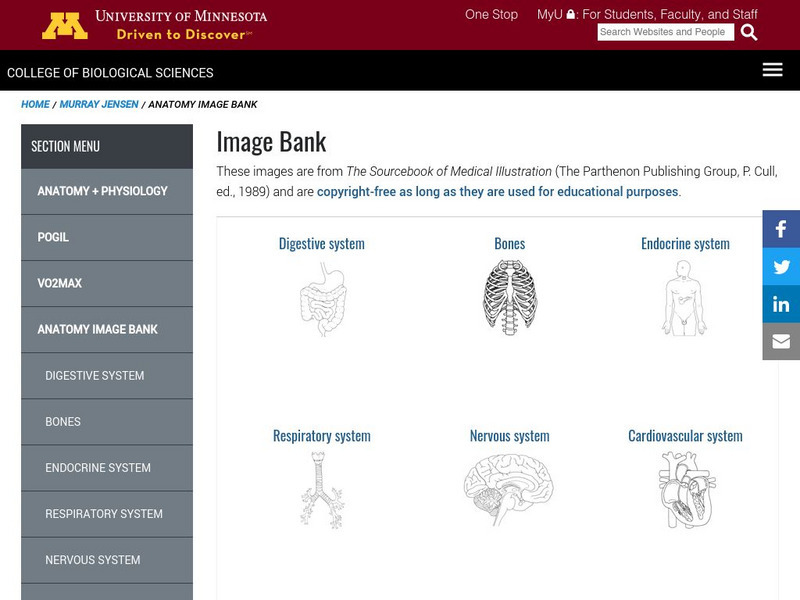
University of Minnesota: Anatomy Image Bank
Hundreds of anatomy diagrams for classroom or medical use. Each image represented is a simple black-and-white professional drawing of a specific part of the human body.
National High Magnetic Field Laboratory
Magnet Academy: Magnetic Resonance Imaging Mri
In MRI, magnetic fields and radio wave pulses combine to get a unique, and medically beneficial, response from your body's hydrogen atoms. Take a peek in this tutorial.
WebMD
Medicine Net: Magnetic Resonance Imaging
This site from MedicineNet.com gives an MRI description, why they're performed, risks, and patient preparation. A thorough explanation of the process is provided.
Massachusetts Institute of Technology
Mit: Invention of the Week: Raymond Damadian: Medical Resonance Scanning Machine
Read about the education and career of Raymond V. Damadian, inventor of the magnetic resonance imaging (MRI), and learn how the MRI has impacted the field of medicine.
National Institutes of Health
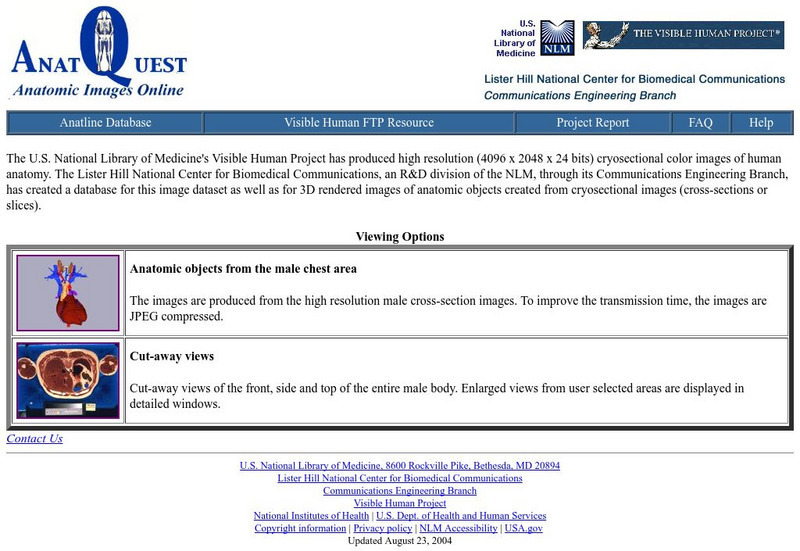
Visible Human Project: Anatomic Images Online
Medical quality 3D images of the male chest area and cut away views of the entire male body. Fantastic for anatomy classes!
Other
Imaios Sas: E Anatomy: Atlas of Human Anatomy
A collection of pictures, videos, medical imaging, and illustrations of human anatomy. A membership is required to view some of the images, while other images can be viewed without one.
Other
Corning Optical Fibers
Corning serves as a leader in optical fiber technology and this website provides information about the uses of optical fibers in medical imaging, communication, etc., as well as industry news and related resources.
National High Magnetic Field Laboratory
Magnet Academy: Paul Lauterbur
Chemist Paul Lauterbur pioneered the use of nuclear magnetic resonance (NMR) for medical imaging. He developed a technique, now known as magnetic resonance imaging (MRI), in the early 1970s that involves the introduction of gradients in...
National High Magnetic Field Laboratory
Magnet Academy: Felix Bloch (1905 1983)
Physicist Felix Bloch developed a non-destructive technique for precisely observing and measuring the magnetic properties of nuclear particles. He called his technique "nuclear induction," but nuclear magnetic resonance (NMR) soon became...
Mocomi & Anibrain Digital Technologies
Mocomi: Mri Scan
Discover what happens during an MRI scan and other interesting facts.
National Institutes of Health
National Library of Medicine: Islamic Culture and the Medical Arts: Al Razi
Short biography of al-Razi, an Islamic "physician learned in philosophy as well as music and alchemy." Includes images of excerpts from his written works.
University of Kansas Medical Center
University of Kansas Medical Center: Cell Structure
Discover microscopic images of various eukaryotic cells, both plant and animal.
University of Kansas Medical Center
University of Kansas Medical Center: Bone
See several specimens of microscopic images of bone cells and tissue.
University of Kansas Medical Center
University of Kansas Medical Center: Female Reproductive System
Examine this collection of microscopic specimen images from the female reproductive system.
University of Kansas Medical Center
University of Kansas Medical Center: Basic Histopathology
These microscopic images of different cells and tissues from the human organs gives an idea of some the pathological processes that occur and the importance of histology.
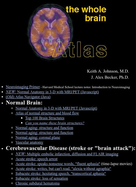
Harvard University
Harvard Medical: The Whole Brain Atlas: Neuroimaging
Hundreds of images of brain structures are here, including normal and diseased brain parts.
Science Daily
Science Daily: Researchers Image Language Recovery After Stroke
This article from ScienceDaily illustrates a recent medical study where researchers at Washington University have identified and imaged language areas of the brain in recovery after stroke.
University of Colorado
University of Colorado: Physics 2000: Cat Scans: Projecting Shadows
This page and the three pages which follow discuss how X-ray technology can be used to produce an image of the human body. Discussion is understandable and highly intriguing. Several interactive animations allow the visitor to explore...
PBS
Pbs Teachers: Scientific American: 21st Century Medicine: Image Guided Surgery
Investigate the science and medical diagnostic uses of magnetic resonance imaging (MRI) technology. Perform experiments to observe how magnetic properties can be induced, and measure the force of temporary magnets.
National Inventors Hall of Fame
National Inventors Hall of Fame: Raymond v. Damadian: Mri
Read about Raymond V. Damadian, inventor of the magnetic resonance imaging (MRI), and learn how the MRI has impacted the field of medicine.
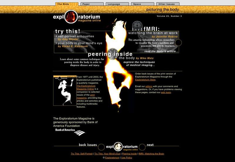
Exploratorium
Exploratorium: Picturing the Body
An online version of articles and activities from the Exploratorium Magazine, Vol. 23, No. 3. This issue looked at how we are able to examine the inside of the human body, what kinds of technology are used, and how each of them is used...


