Museum of Science
Mo S: Image Gallery of the Scanning Electron Microscope
At this resource you'll find everything from a mosquito head to a fly foot, from a dentist's drill to toilet paper! These and just about everything in between as seen through a scanning electron microscope.
Science Struck
Science Struck: Light Microscope vs Electron Microscope: A Comparison
Looks at the differences and similarities between a light microscope and an electron microscope. Discusses their structure, how images are formed, their resolution and magnification, how color is manifested, their portability, cost, and...
University of Illinois
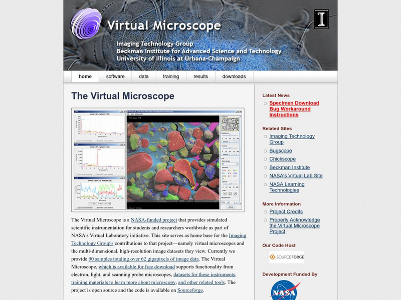
University of Illinois Urbana Champaign: Imaging Technology Group: Virtual Microscope
After a free download, the Virtual Microscope simulates the experience of using an electron, light and scanning probe microscope. The microscope basics are also explained through a series of animations.
CK-12 Foundation
Ck 12: Plix: Microscopes
[Free Registration/Login Required] Focus a virtual microscope and learn about the images microscopes produce on this site. Also included is a short quiz on microscopes.
TeachEngineering
Teach Engineering: Imaging Dna Structure
Students are introduced to the latest imaging methods used to visualize molecular structures and the method of electrophoresis that is used to identify and compare genetic code (DNA). Students should already have basic knowledge of...
Exploratorium
Exploratorium: Microscope Imaging Station: Cell Development
This interesting site provides videos and photos of cells going through the process of cell division. Observe Zebrafish development and Sea urchin embryo cell division in video format.
Other
Science Stock Photography
This site has numerous pictures of microscopic images. It also has a search engine so you can find specific objects. Images were captured with light microscopes, as well as electron microscopes.
Exploratorium
Exploratorium: Microscope Imaging Station: Cell Motility
This interesting site explains how cells move and provides videos depicting the movement of various human and animal cells.
Exploratorium
Exploratorium: Microscope Imaging Station: Blood Cells
This colorful site provides a gallery of blood cell photos. You can observe human white blood cells, human red blood cells, sheep red blood cells, and zebrafish red blood cells. As an added bonus, you can enlarge the red blood cell...
Microscopy UK
Microscopy Uk: An Introduction to Microscopy
An introduction to the world of the microscope. Includes information about the instrument and the subjects you can study through the microscope.
Museum of Science
Museum of Science: Scanning Electron Microscope (Sem)
View a video or a click-through a slideshow to find out how the SEM works. In addition, find an interesting gallery of magnified images captured with an SEM, links to related sites, and a teacher resource section.
Florida State University
Florida State University: Magnification Module
Explore the effect of increasing the magnification on a microscope when viewing various samples such as onion root mitosis.
Florida State University
Florida State University: Translational Microscopy
With this interactive tutorial, you can view cholesterol, pesticide, ibuprofen, and other samples under a microscope. You can adjust the focus, intensity, and zoom in on the object. Requires Java.
Other
Scimat: Foods Under the Microscope
A technical site that describes different types of microscopes and delves into the chemical makeup of milk, yogurt, various various types of microorganisms. Impressive pictures supplement the text. Links to images of microorganisms.
Other
Xtalent Art Gallery: Nanoworld Image Gallery
Microscopic gallery of various organisms and objects.
Florida State University
Florida State University: Molecular Expressions: Basic Concepts in Optical Microscopy
This site explores the microscope in great detail. Content includes a focus on the microscope's basic functions (including a general diagram), the history of the microscope, each feature of the microscope, differing objectives,...
Georgia State University
Georgia State University: Hyper Physics: Thin Lens Equation
This is an informative site from Georgia State University. It gives a discussion of the thin lens equation and an illustration of its use in determining the image distance based upon the object distance and the focal length.
Arizona State University
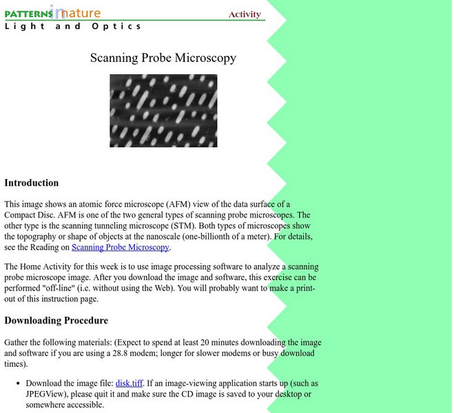
Arizona State University/spm Home Activity
Investigate a SPM image at home with this activity of the week. Links back to more info about SPM's.
Other
Southern Microscope Service's Home Page
A series of pages explaining how microscopes work, the parts of a microscopes, the techniques of using a microscope, and other useful information.
Other
University of Muenchen: Scanning Probe Microscopy Group
Scroll down to research to find images of scanning tunneling and atomic force microscopes.
Other
University of Western Australia: Blue Histology
An excellent, in-depth examination of the integumentary system and its components. Includes many color microscopic images of these structures and more.
Other
University of Western Australia: Integumentary System
A resource for advanced anatomy and physiology classes. It contains information on the integumentary system, the epidermis, the dermis, hair, sebaceous glands, and sweat glands. Microscopic images are included.
Other
National Institute for Basic Biology: The Plant Organelles World
This web resource provides basic information about structure and function of plant organelles through a large interactive cell drawing, as well as many opportunities to explore further. Links lead the learner to several short movies of...


